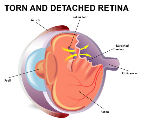
Retinal Detachment
Retinal Detachment – is a disorder of the eye in which the retina, the inner light sensitive layer of the eye separates from the middle layer of the eye. Symptoms include an increase in the number floaters, flashes of light, and partial loss of side vision. This is often described as a curtain over part of the field of vision.
Cause of Retinal Detachment
A clear gel called vitreous fills the back of the eye. As we get older, the vitreous eventually pulls away from its attachment to the retina at the back of the eye, usually without causing problems. Sometimes, though the vitreous pulls hard enough to tear the retina in one or more places. Fluid may pass through the retinal tear blistering the retina off the back of the eye, much as wallpaper can peel off a wall.
Conditions that can increase the chances of having a retinal detachment:
- Nearsightedness
- Previous eye surgery
- Glaucoma
- Severe injury or trauma to the eye
- Previous retinal detachment in your other eye
- Family history of retinal detachments
Treatments for Retinal Tears
Retinal tears can be treated in the office. We typically do this the same day we diagnose the tear, because we can avoid the development of an RD if we treat the retinal tear in time. The main treatment for a tear is laser surgery, but at times we have to do a freezing cryotherapy treatment as well, depending on the size and location of the tear.
Treatments for Retinal Detachments
There are multiple surgical treatment options for retinal detachments.
- Scleral Buckle – This treatment is performed in an operating room under either a local or general anesthetic. It involves placing a flexible band around the eye to counteract the force pulling the retina out of place.
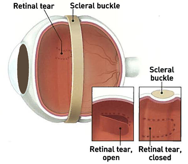
Detached Retina: Scleral Buckle
- Pneumatic Retinopexy – This treatment can be performed either in an operating room or in our office. In this procedure,re a gas bubble is injected into the vitreous space inside the eye in combination with laser or cryotherapy. The purpose of the gas bubble is to push the retinal tear closed against the back wall of the eye. You will be asked to maintain a certain head position for several days. The gas bubble will gradually disappear.
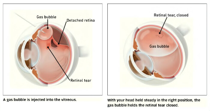
Detached Retina: Pneumatic Retinopexy
- Vitrectomy – This treatment is performed in an operating room under either local or general anesthesia. The vitreous gel, which is pulling on the retina, is removed from the eye and usually replaced with either a gas or oil bubble to keep the retina in place. Your body’s fluids will gradually replace a gas bubble. An oil bubble has to be removed from the eye at a later date with another surgical procedure. Sometimes a Vitrectomy is combined with a Scleral Buckle.
Detached Retina: Vitrectomy
It is important to seek treatment as soon as your symptoms occur to increase your chances of preserving vision.